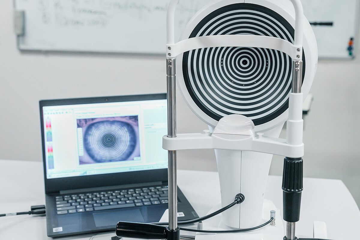
16:00 Renārs Trukša ( Latvijas Universitāte) Promocijas darba priekšaizstāvēšana Datorizētas krāsu redzes testu platformas izstrāde.
Renāra Trukša promocijas darbs ir veltīts krāsu redzes problemātikai. Promocijas darba ietvaros ir izstrādāts modelis, lai paredzētu cilvēku sarkani-zaļajiem krāsu redzes deficītiem sniegumu krāsu sakārtošanas testos. Izstrādātais modelis korekti paredz cilvēku ar sarkani-zaļiem krāsu redzes deficītiem sniegumu D15 testā atkarībā no deficīta izteiktības pakāpes. Modelis apstiprina iepriekš zināmas hipotēzes par krāsu redzi kā arī sniedz ieskatu par krāsu izšķirtspējas atšķirībām cilvēkiem ar krāsu redzes deficītiem. Doktora disertācijas ietvaros ir izstrādāts datorizēts krāsu redzes tests, kas ietver divu tipu stimulus – dinamisku un statisku. Izstrādātais krāsu redzes tests sniedz iespēju konstatēt normai atbilstošu krāsu redzi, kā arī identificēt sarkani-zaļos krāsu redzes deficītu gadījumus. Pētījuma ievaros izvērtēta izstrādātas metodes efektivitāte salīdzinot to ar tādiem krāsu redzes testiem kā HRR pseidoizohromatisko plašu tests, FM100 krāsu sakārtošanas tests, CAD (Colour Assesment and Diagnosis test) datorizētais krāsu redzes tests un anomaloskopa tests. Promocijas darba ietvaros izvērtēts datorizētā testa potenciālais pielietojums praksē, kā arī izvērtēta jau esošo krāsu redzes testu un to kombināciju efektivitāte, lai identificētu krāsu redzes deficītus.
17:00 Elena Salobrar García Martín (Universidad Complutense de Madrid) OCT and OCT-A: image capture and interpretation for optometrists. Application in neurodegenerative diseases: Alzheimer's Disease.
In Vision Sciences, Optical Coherence Tomography (OCT) is a non-invasive and inexpensive technique that uses a non-coherent beam of light to allow us to see the retina, including the macula and optic disc, and analyze the neurons that populate the retina with a resolution of microns. This technique enables us to segment the layers of the retina and, due to its stratified structure, determine the thickness of the retina in each of its layers and identify the neuronal population that is being affected. Furthermore, analysis of the peripapillary retina allows us to determine the thickness of the fiber layer, formed by the axons of the ganglion cells of the retina that project to the midbrain. This technique allows us to explore the neurons of the retina in a non-invasive manner and provides valuable information in the study of ocular pathology.
In this talk, we will learn about the different OCT devices available on the market and their peculiarities. We will understand how to interpret the different images provided by OCTs from a histological perspective, and learn about the different artifacts that these devices can provide in order to improve capture quality. Additionally, as changes in the retina have been described in different neurodegenerative diseases, we will use the knowledge acquired to understand what happens in the retina of patients with Alzheimer's disease and what findings we can expect to see in an OCT exam of these patients.
Redzes zinātnes doktorantūras skolas nodarbība notiks LU Dabas mājā Jelgavas ielā 1 - 223. telpā plkst. 16:00 un tiešsaistē BBB platformā. Pieslēgšanās links.
Nodarbībai jāpiesakās līdz 12. aprīlim, aizpildot anketu.
 organizētajā Redzes zinātnes doktorantūras skolā ar promocijas darba priekšaizstāvēšanu uzstāsies Lekt. Renārs Trukša un vieslektore no Spānijas. )